Your knee pain, explained simply
No medical jargon. Just what's happening inside your knee, what your options are — surgical and non-surgical — and what to expect at each stage.
What is knee osteoarthritis?
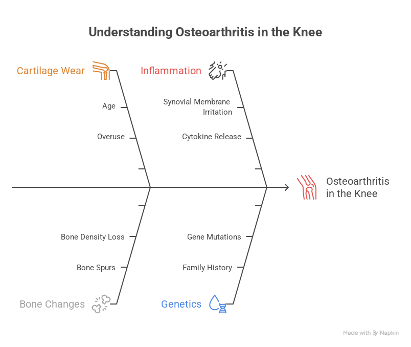
Your knee has a smooth cushion of cartilage that lets the bones glide against each other painlessly. Osteoarthritis (OA) means this cushion is thinning out. As it wears down, the bones start rubbing closer together — causing the stiffness, swelling, and pain you feel, especially when climbing stairs, sitting cross-legged, or standing up after rest.
It's not something that happened overnight, and it's not something you caused. It's a gradual, mechanical wearing-down — most common after 45, faster in overweight or previously injured knees.
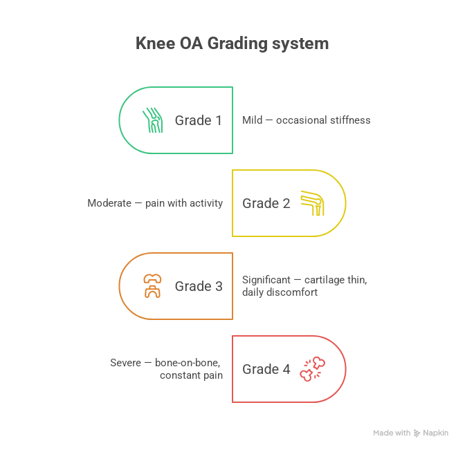
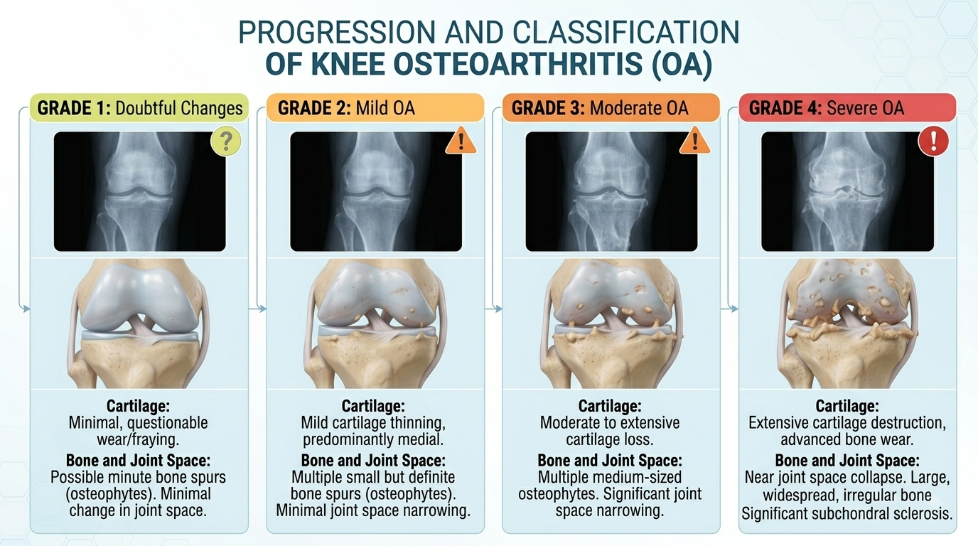
The 4 grades of knee OA
Your X-ray tells us how much cartilage cushion is left. This decides which treatments make sense for you — most knees do not need surgery.
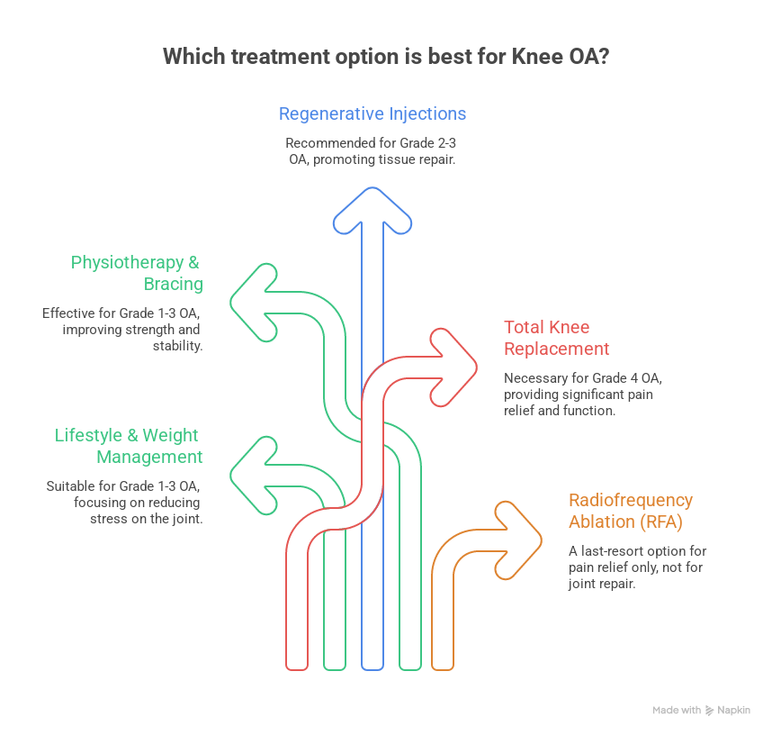
The treatment ladder
We always start from the top of this ladder and only move down if you need to. Surgery is the last step — not the first.
Lifestyle & weight management
Reduces load on the joint. Often enough on its own for mild OA.
Physiotherapy & bracing
Strengthens the muscles around the knee to take pressure off the worn cartilage.
Regenerative injections PRP · BMAC · MFAT
Your own blood or bone-marrow-derived cells are concentrated and injected into the knee to reduce inflammation and support the joint's own repair — without surgery. Effective only while some cartilage cushion remains, up to Grade 2–3.
Allogeneic stem cell therapy DGCI Approved
Lab-processed donor stem cells for patients who need a stronger regenerative option than their own body can provide. Like other injectables, this is not indicated for Grade 4 (bone-on-bone) knees.
Total Knee Replacement (Robotic-assisted)
Considered only when the cartilage is gone and non-surgical options have stopped giving relief.
How a total knee replacement is performed
In a Total Knee Replacement (TKR), the worn cartilage and a thin layer of damaged bone are removed and replaced with a smooth metal-and-plastic implant that recreates a pain-free, well-aligned joint.
Planning
Robotic/CT-based mapping of your exact knee shape, so the implant is sized and aligned specifically to you.
Access
A small incision over the front of the knee opens the joint.
Resurfacing
The damaged bone-and-cartilage surfaces of the thigh bone and shin bone are precisely trimmed — robotic guidance keeps this accurate to within a millimetre.
Implant placement
Metal caps are fixed onto the bone ends with a smooth plastic spacer between them, recreating the joint surface.
Movement check & closure
The knee is bent and tested through its full range before closing, to confirm smooth, stable movement.
Pros
- Reliable, long-lasting pain relief
- Restores ability to walk, climb stairs, sit normally
- Robotic precision improves implant alignment & longevity
- Most patients walk the same day or next day
Cons
- Major surgery — needs anaesthesia and hospital stay
- 4–6 week recovery before returning to normal routine
- Implant has a functional lifespan (typically 20+ years)
- Physiotherapy commitment is essential for best results
What if you're not fit for surgery?
A small number of Grade 4 patients have other health conditions that make surgery too risky. For them, and only them, Radiofrequency Ablation (RFA) is an option.
RFA does not repair or regrow your joint. It works by numbing the nerves that carry pain signals from the knee. This means it does not fix the underlying problem — it only silences the pain — and it is considered a last resort, not a routine alternative to surgery or a step to take before trying surgery.
Your recovery, week by week
Physiotherapy begins within hours. Most patients stand and take a few steps the same or next day.
Hospital discharge once you can walk safely with a walker and manage stairs.
Stitches/staples removed. Walking indoors without support for many patients.
Most daily activities resume — walking outdoors, driving (once reflexes are confirmed safe).
Full bend and strength continue improving. Most patients feel "normal" in daily life.
Peak recovery — walking, stairs, travel, and most sports/activities without knee pain.
Patients often ask
Can I avoid surgery altogether?
If you're Grade 1–3, very often yes — with weight management, physiotherapy, and regenerative injections. Grade 4 (bone-on-bone) is where surgery becomes the most reliable long-term fix.
Is robotic knee replacement different from a normal one?
The surgery achieves the same goal, but robotic guidance maps your knee precisely beforehand and keeps bone-cutting accurate to within a millimetre — which improves implant alignment and how naturally the knee moves afterward.
How painful is the recovery?
Pain is managed with medication from day one, and most patients are surprised at how quickly they're up and walking. Physiotherapy is uncomfortable but not unbearable — and essential to a good outcome.
Will PRP or stem cell therapy work for me?
It depends on your grade. These work best in Grade 2–3 knees that still have cartilage to support. An X-ray/consultation is the only way to know which ladder step is right for you.
Not sure which stage you're at?
Share your X-ray or symptoms on WhatsApp and Dr. Sumit's team will guide you on the right next step.
Message us on WhatsAppThis guide is for general patient education and does not replace an in-person clinical evaluation.
← Back to all handbooks